Research Article
Detection of Levodopa in Rat Plasma by Capillary Electrophoresis
Elizabete Lourenço da Costa*, Antonio José Calixto de SOUZA, Alessandra de Sá CUSTODY and Débora Mendoça Rabelo CARVALHO
Corresponding Author: Elizabete Lourenço da Costa, Anhembi Morumbi University, Almeida Lima St. 1134, São Paulo - SP, Brazil.
Received: December 06, 2022; Revised: December 11, 2022; Accepted: December 24, 2022 Available Online: January 11, 2023
Citation: da Costa EL, de SOUZA AJC, de Sá CUSTODY A & CARVALHO DMR. (2023) Detection of Levodopa in Rat Plasma by Capillary Electrophoresis. J Pharm Drug Res, 6(2): 701-703.
Copyrights: ©2023 da Costa EL, de SOUZA AJC, de Sá CUSTODY A & CARVALHO DMR. This is an open-access article distributed under the terms of the Creative Commons Attribution License, which permits unrestricted use, distribution, and reproduction in any medium, provided the original author and source are credited.
Share :
  • 455

    Views & Citations
  • 10

    Likes & Shares
L-dopa is one of the most used drugs for the treatment of Parkinson’s disease, but its absorption can be decreased by food interference. In this work we show that capillary electrophoresis (CE) can be a useful method for clinical, pharmacological or therapeutic L-dopa bioavailability studies. The experiment consisted in a sublethal oral administration to Wistar rats, after 2h blood samples were collected, plasma proteins were precipitate with trichloroacetic acid addiction; the solution was filtered and injected in silica capillary. L-dopa was efficiently separated and quantified by measuring the absorbance at 236nm. The CE step is rapid and can be performed in about 7 min.

Keywords: L-dopa, Parkinson’s disease, Capillary electrophoresis, Method
INTRODUCTION

Parkinson's disease (PD) affects more than 1 percent of persons aged 65 years or older. PD is associated with resting tremor, cogwheel rigidity, and bradykinesia. Others non-motor manifestations, like dementia, constipation, sleep disorders, and erectile dysfunction, are common [1].

L-dopa has been used as a prodrug of dopamine in the treatment of PD; its metabolism is a complex process, with many pathways, with a range of inter- and intra-patient variation of oral bioavailability, due to its interaction with foods, attributed to amino acid transport competition [2].

L-dopa is the most effective anti-Parkinson in agent, but the most of patients with PD can be expected to develop dyskinesias, or involuntary movements, within 5 years of starting therapy. Much progress has been made in elucidating the causation and pathogenesis of PD; we know that PD is complex, stimulated by both genetics and environmental factors [3].

The L-dopa plasma concentration can be a fundamental parameter for measuring the oral bioavailability drug or the drug therapy efficacy in clinical studies, and provide the better adjustment of treatment conditions, however in case of patients requiring high doses is common the appearance of motor complications that consists in wearing-off fluctuations and abnormal involuntary movements. So investigating serum levels can bring a better evaluation between the symptoms and the dopaminergic medications dose [4].

There are many methods to determination of L-dopa, including high-performance chromatography, thin layer chromatography, fluorescence and colorimetric methods [5,6] but must of them are carried out for plant matrices. Here we describe that capillary electrophoresis (CE) can be used also, to determine L-dopa in serum using TCA precipitating the plasma proteins, with the advantage of using small sample volume, and low amount of disposal generated, as compared with chromatographic methods.

MATERIAL AND METHODS

The experiments were performed with male Wistar rats (n = 6), weighing about 250 to 260g each, allowed free access to water and food. Animals were housed in a 12h light/dark cycle, with 25±1°C constant temperature environment.
In order to evaluate the L-dopa absorption, the animals received an intraperitoneal (i.p.) sublethal dose of commercial L-dopa associated to carbidopa (3000mg/kg wt).

After 2h, the blood was collected under anesthesia by via cardiac punction, and centrifuged (200xg/10 min), plasma proteins were precipitate with trichloro acetic acid (TCA). The ethical aspects were respected in accordance with the general guidelines established by the Brazilian College of Animal Experimentation (COBEA), and the Helsinki Declaration was followed throughout the study.

The determination of plasma L-dopa was performed by a capillary electrophoresis system (HP-3DCE Agilent, Waldron, Germany); the samples were filtered in an acetate cellulose membrane (0, 22mm) and injected directly on silica capillary. The injections were in the hydrodynamic mode in silica capillary (52cm x 75mm ID; Agilent), and the pressure was 50mBar. The run voltage applied was 10kV. The runs were performed in borate buffer 50mM, pH 9.0. L-dopa was detected at 236 nm using a Diode Array Detector. Pure L-dopa was used as standard to compare migration time and UV spectra.

RESULTS AND DISCUSSION

CE is well suited for diagnostic analysis, been used to determination of forensic analysis, due its applicability includes analysis of drugs, serum proteins, hemoglobin variants, and others molecules [6].

The importance of measuring serum L-dopa consists in the possibility to monitor the PD-patient that can present a poor absorption, depending of his food habits, and the time of the disease progression.

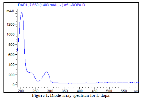
The maximum absorption of L-dopa is about 200nm, although this wavelength has more possibility of interferences from serum peptides, small protein, or other unknown compounds. The Figure 1 shows the L-dopa spectra, with no spectral features in the visible region, but with two others peaks at 236 and 285nm that can be used during analysis to decrease the interfering possibility.
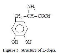
Using a run buffer of 50mM borate of pH 9.0 facilitated a rapid analysis time, due its negative charge net of unproton carboxylic group, with migration time of 5.4 min (Figure 2). The L-dopa structure is shown in Figure 3.
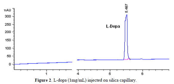

A linear detector response was obtained when decreasing concentrations of L-dopa were injected in the silica capillary.

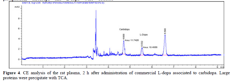
Figure 4 show a rat plasma CE profile after protein precipitation, carbidopa and L-dopa were detected separately. The L-dopa spectrum obtained was consistent with the previously found after standard injection (Figure 1). The L-dopa plasma concentration average was 5.44±0.37mg/mL.
Tesoro [6] made an overview of methods to extract and analyze L-Dopa, including an electrophoresis capillary method; however the measurements are done in conditions that extract this analyte since plant matrice.

The method preliminary developed in this study is simple, rapid, and suitable for the quantification of L-dopa or carbidopa in plasma or other biological fluid samples from clinical, pharmacological or therapeutic studies. In additional, diode array detection can reduce doubts about analytes nature, because in L-dopa oxidation several intermediate products are formed.

ACKNOWLEDMENTS

The authors tank Dr. Jaime Amaya-Farfán, Faculty of Food Engineering of the State University of Campinas-SP, Brazil, for provide the capillary electrophoresis apparatus.


  1. Cenci MA, Crossman AR (2018) Animal models of L-dopa-induced dyskinesia in Parkinson's disease. Mov Disord 33(6): 889-899.
  2. Fabbrini A, Guerra A (2021) Pathophysiological mechanisms and experimental pharmacotherapy for L-dopa-induced dyskinesia. J Exp Pharmacol 29(13): 469-485.
  3. Jiang R, Yang J, Mei S, Zhao Z (2022) Determination of levodopa by chromatography-based methods in biological samples: A review. Anal Sci 38: 1009-1017.
  4. Pang SYY, Ho PWL, Liu HF, Leung CT, Li L, et al. (2019) The interplay of aging, genetics and environmental factors in the pathogenesis of Parkinson’s disease. Transl Neurodegener 8: 23.
  5. Puris E, Gynther M, Auriola S, Huttunen KM (2020) L-Type amino acid transporter 1 as a target for drug delivery. Pharm Res 37: 88.
  6. Tesoro C, Lelario F, Ciriello R, Bianco G, Capua DA, et al. (2022) An overview of methods for L-dopa extraction and analytical determination in plant matrices. Separations 9: