Journal of Oral Health and Dentistry (ISSN: 2638-499X)

Mini-Review
Effect of Thickness on the Fracture Resistance of Ceramic Partial Restorations: A Review
4084
Views & Citations3084
Likes & Shares
This publication describes the effect of thickness and type of material on the resistance fracture of ultra-thin ceramic restoration. The restorative phase of the treatment should not cause additional damage of the residual tooth structure. Ultrathin restorations (veneers, onlays, inlays) are considered as an alternative to traditional onlays and complete crowns. the technical aspects required for the success and the good prognosis of those new restorative design based on the control of tooth preparation with diagnostic wax-up, provisionalization, and the use of CAD-CAM technology.
Keywords: Ultrathin, Restoration, Ceramics, Thickness, Fracture
INTRODUCTION
The dental enamel is designed to withstand a lifetime. Her progressive reduction is biological consequence of advancing age. The loss of tissue may be due to the action of acidic foods, gastroesophageal reflux disease medications, and the reduction of salivary flow [1-3].
There are now many new protocols for a new concept of ultrathin and no pre ceramic restorations. Ceramic veneers and inlays, onlays and overlays are frequently presented as the major class of clinical conservative modalities [4].
CAD/CAM technology became popular during the last decade for the conception and fabrication of restorations. Different materials are supplied in the form of blocks that are milled to obtain the restorations [3].
Among ceramic CAD/CAM materials, lithium disilicate have recently expanded their indications to include ultra-thin restorations, with promising results. Recently, hybrid ceramic (vita Enamic) has been developed to allow faster milling of the ceramic block as ultrathin restorations with good mechanical behavior and good prognosis.
Due to the importance of dental tissue preservation, it’s important to evaluate the fracture resistance of reduced thickness materials made with different restorative materials [4,5].
EFFECT OF THICKNESS
It’s very notable how well patients presenting with tooth fracture, moderate to severe loss of tooth structure when ultra-thin restorations are proposed to them, both economic and biological costs are significantly lower compared to traditional and more invasive approaches. Minimal thickness can be used were in the Table 1 [1,2,6-8].
The possibility of making ultrathin (0.3 mm-0.6 mm) ceramic restoration allows for a more conservative preparation with minimal wear to the tooth structure.
It’s believed that these positive and promising results are due in part to the adhesive luting technique, dental substrate and restorative material [9].
According to the study of Nordahl [10], comparing five thickness (0.3-0.5-0.7-1-1.5) for high-translucent (HTZ) and low-translucent (LTZ) zirconia restorations and glass ceramic (LDS) crowns. The lowest recorded load at fracture within & mm groups was 634 N, and 550N for the Y-TZP groups at thickness of 0.5mm. Compared to the forces measured during mastication (approximately 5 to 364N); the results suggest the possibility to reduce restorations
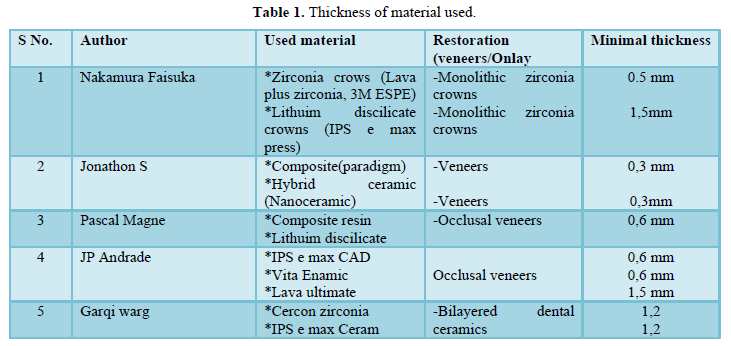
On the other hand, the study of JP Andrade Showed that the fracture resistance was significantly higher at a thickness of 1,5 mm compared to a thickness of 0,6 mm for veneers made of lava ultimate and vita Enamic.
Manufactures of lava Ultimate, vita 3namic and IPS e.max CAD, affirmed that restorations with a minimum thickness of 1.5 mm on the occlusal surface of posterior teeth will support masticatory loads. Nevertheless, other studies showed that it is possible to treat severe erosive lesions or loss of wear on posterior teeth with ultrathin (0.5-1mm) caeramic and composite resin materials [12].
The study of JP Andrade, evaluated 0,6 mm of thickness (veneers), which are considered ultrathin restorations. On the other hand, the study of Egbert et all, shows a fracture resistance of occlusal veneers with a 0.3 mm using Paradigm MZ 100, Vita Enamic and lava Ultimate; and found promising fracture resistances. Hence, it seems that the use of use of thickness smaller than 0,6 mm could be used with good prognosis [3].
THE LUTING MATERIAL AND PROTOCOL
Ceramic indirect restoration luted by the adhesive luting technique provided better fracture resistance than conventional luting technique. Hence, the use of adhesive restorations has been recommended for reinforcing the remaining dental structure because It allows intimate contact between the dental substrate, luting agent, and ceramic material, therefore occlusal forces are dissipated through the root of tooth, periodontal ligament, and alveolar bone [5,8].
According to many studies, associating hydrofluoric acid with silane was the most effective surface treatment with which to potentiate the bond between the ceramic and the adhesive material [2,5,7].
The silane enhances the chemical bond between the silicon-containing materials and the resinous material used for luting.
THE TYPE OF MATERIAL
The study of Heck [11] showed that IPS e max CAD and lava ultimate should be preferred to IPS Empress CAD for the treatment of occlusal tooth loss with ultrathin restoration, whether this result is due to the viscoelastic proprieties of the composite material. In another study, Johansson [12], compared fracture resistance of monolithic zirconia and monolithic lithium disilicate after cyclic loading and thermos-cycling. they reported higher strength for zirconia restorations with the same occlusal thickness (0.5mm and 1mm). According to Nordhal [10] Ceramic materials, such as glass ceramics and zirconia show a greater scatter in fracture strength compared to other material such as metal. This result calls for special factor approach when indicating reduced ceramic restoration [5,7-9].
CONCLUSION
Ultrathin restorations (inlay, onlays, veneers.) appear to be a promising restorative procedure in posterior and anterior teeth.
The feasibility of their application depends on their fabrication options and fracture properties. Recent advances in technology and materials are offering new options for good treatment [13,14].
- Nakamura K, Harada A, Inagaki R, Kanno T, Niwano Y, et al. (2015) Fracture strength of ceramic monolithic crown systems of different thickness. Acta Odontol Scand 73(8): 1-7.
- Wang G, Zhang S, Bian C, Kong H (2015) Effect of thickness ratio on load-bearing capacity of bilayered dental ceramics. J Prosthodont 24(1): 17-24.
- Schilchting LH, Maia HP, Baratieri LN, Magne P (2011) Novel-design ultra-thin CAD/CAM composite resin and ceramic occlusal veneers for the treatment of severe dental erosion. J Prosthet Dent 105(4): 217-226.
- Johnson AC, Verluis A, Tantbirojen D, Ahuja S (2014) Fracture strength of CAD/CAM composite and composite-ceramic occlusal veneers. J Prosthodont Res 58(2): 107-114.
- Magne P, Schilchting LH, Maia HP, Baratieri LN (2010) In vitro fatigue of CAD/CAM composite resin and ceramic posterior occlusal veneers. J Prosthet Dent 104(3): 149-157.
- Egbert JS, Johnson AC, Tantbirojn D, Versluis A (2015) Fracture strength of ultrathin occlusal veneer restorations made from CAD/CAM composite or hybrid ceramic materials. Int J Oral Sci 12(2): 1348-8643.
- Andrade JP, Stona D, Bittencourt HR, Borges GA, LH Burnett LH Jr, et al. (2018) Effect of different computer-aided design/computer-aided Manufacturing (CAD/CAM) materials and thicknesses on the fracture resistance of occlusal veneers. Oper Dent 43(5): 539-548.
- Magne P, Stanley K, Schlichting LH (2012) Modeling of ultrathin occlusal veneers. Dent Mater 28(7): 777-782.
- Resende TH, KR Reis KR, Schlichting LH, Magne P (2018) Ultrathin CAD-CAM ceramic occlusal veneers and anterior bilaminar veneers for the treatment of moderate dental biocorrosion: A 1.5-Year follow-up. Oper Dent 43(4): 337-346.
- Nordahl N, von Steyern PV, Larsson C (2015) Fracture strength of ceramic monolithic crown systems of different thickness. J Oral Sci 57(3): 255-261.
- Heck K, Paterno H, Lederer A, Litzenburger F, Hickel R, et al. (2019) Karl-Heinz Kunzelmann Fatigue resistance of ultrathin CAD/CAM ceramic and nanoceramic composite occlusal veneers. Dent Mater 35(10): 1370-1377.
- Johansson C, Kmet G, Rivera J, Larsson C, Von Steyern PV (2014) Fracture strength of monolithic all-ceramic crowns made of high translucent yttrium oxide-stabilized zirconium dioxide compared to porcelain-veneered crowns and lithium disilicate crowns. Acta Odontol Scand 72: 145-153.
- Dirxen C, Blunck U, Preisser S (2013) Clinical performances of a new biomimetic double network material Open Dent J 7(6): 118-122.
- Egbert JS, Johnson AC, Tantbirojn D, Verluis A (2015) Fracture strength of ultrathin occlusal restorations made from CAD/CAM composite or hybrid ceramic materials. Oral Sci Int 12(2): 53-58.
QUICK LINKS
- SUBMIT MANUSCRIPT
- RECOMMEND THE JOURNAL
-
SUBSCRIBE FOR ALERTS
RELATED JOURNALS
- Chemotherapy Research Journal (ISSN:2642-0236)
- Journal of Infectious Diseases and Research (ISSN: 2688-6537)
- Journal of Cancer Science and Treatment (ISSN:2641-7472)
- Journal of Nursing and Occupational Health (ISSN: 2640-0845)
- Advance Research on Endocrinology and Metabolism (ISSN: 2689-8209)
- International Journal of Medical and Clinical Imaging (ISSN:2573-1084)
- International Journal of Radiography Imaging & Radiation Therapy (ISSN:2642-0392)


