BioMed Research Journal (ISSN:2578-8892)

Research Article
Estimation of Mechanistic Biomarkers in Multiple Organs after Administration of Bio field Energy Treated Proprietary Test Formulation on L-NAME and High Fat Diet-Induced Cardiovascular Disorders in Sprague Dawley Rats
12920
Views & Citations11920
Likes & Shares
Cardiovascular disorders (CVDs) are one of the most leading cause of death worldwide. Study objective was to evaluate the impact of the Biofield Energy Treated/Blessed novel Proprietary Test Formulation and Biofield Energy Healing Treatment/Blessing per se to the animals on NG-nitro-L-arginine methyl ester A hydrochloride (L-NAME) and high fat diet (HFD)-induced cardiovascular model in Sprague Dawley rats using various functional organ (liver, heart, kidney, brain and muscles) biomarkers. The functional biomarkers like vitamin D receptor (VDR), telomerase enzyme, and citrate synthase using ELISA assay for the assessment of overall health benefits. Each constituent of the test formulation was divided into two parts; one part was known as the untreated test formulation. The other part of each ingredient and of animals (three group) were received Biofield Energy Healing Treatment/Blessing remotely for ~3 minutes by Mr. Mahendra Kumar Trivedi, a renowned spiritual leader. The result showed that the level of VDR expression in the liver tissue was significantly increased by 25.12% and 30.03% in the G5 (L-NAME + HFD + the Biofield Energy Treated test formulation) and G6 (L-NAME + HFD + Biofield Energy Treatment per se to animals from day -15) groups, respectively, as compared to the (L-NAME + HFD + untreated test formulation) group (G4). Moreover, the level of VDR in the kidney tissue was also increased by 21.08%, 31.30%, 15.12%, 17.12%, and 15.06% in the G5, G6, G7 (L-NAME + HFD + the Biofield Treated/Blessed test formulation from day -15), G8 (L-NAME + HFD + Biofield Energy Treatment/Blessing per se plus the Biofield Energy Treated/Blessed test formulation from day -15), and G9 (L-NAME + HFD + Biofield Energy Blessed animals with untreated test formulation) groups, respectively as compared to the disease control (G2) group. Additionally, the expression of VDR in the heart tissue was significantly increased by 25.23%, 25.45%, and 19.42% in the G5, G6, and G8 groups, respectively as compared to the G2 group. Further, the level of telomerase enzyme in the brain tissue was increased by 88.67%, 78.59%, 120.29%, 93.24%, and 87.63% in the G5, G6, G7, G8, and G9 groups, respectively than G2 group. Further, the level of citrate synthase was increased marginally in the treatment groups as compared to the disease control and untreated test formulation group. Overall, data suggested a significant improvement of VDR expression and telomerase enzyme activity in different tissue homogenate, Thus, the Biofield Energy Treated test formulation and Biofield Energy Treatment per se along with preventive measure on the animals that might be beneficial various types of cardiovascular disorders and overall health. Therefore, the study outcomes showed the significant slowdown the oxidative stress-induced cardiovascular disorders and its related symptoms in the preventive treatment groups (viz. G6, G7, G8, and G9) compared to disease control group.
Keywords: Biofield Treatment, Mitochondrial Assay, VDR Assay, Telomerase assay, The Trivedi Effect®, ELISA, High Fat Diet, Cardiovascular Disorders
INTRODUCTION
Cardiovascular disorders (CVDs) are one of the most leading cause of death worldwide [1]. World health organization (WHO) reported that approximately 17.9 million people died due to CVDs per year [2]. Most aged peoples faced some sort of memory loss, which is supposed to be related with various brain aging biomarkers. According to the WHO, aged population number has been significantly increasing and would be doubled in 2050 [3,4]. The mechanistic biomarker is basically used in the clinical diagnosis of symptomatic disease such as, the presence of infectious diseases could be detected by analyzing the antibodies directed against specific pathogens (e.g., HIV or hepatitis virus); diagnosis of certain cancers by detecting the specific genetic aberrations (including myelodysplastic syndrome and chronic myelocytic leukemia) [5]. In animals, the vitamin D deficiency has been reported to result in the cardiovascular consequence i.e., hypertension. Some research studies on rats reported that the deficiency in vitamin D receptor (VDR) may develop hypertension in them. It could be associated with the activation of the renin-angiotensin-aldosterone system, as vitamin D acts as a negative regulator of renin synthesis [6]. Telomerase is a reverse transcriptase enzyme, that can restore DNA known as telomeres, otherwise shortened the telomere length, during cell division via mitosis. In humans, patients with atherosclerotic diseases and cardiovascular risk factors are more prone to shorter leukocyte telomere length. Besides, telomerase plays a vital role in regulating tissue repairs in heart diseases. Hence, heart tissue is selected in this study for the estimation of telomerase enzyme level in heart homogenate [7,8]. Brain post-mortem studies revealed that no difference in telomere length is observed in depression. Telomere length is highly dependent on the telomerase enzyme activity. In brain tissue telomere shortening has been observed in grey matter. Hence, brain tissue is also selected in this study for the estimation of telomerase enzyme level [9]. Several biochemical measures of mitochondrial components are used as biomarkers of mitochondrial content and muscle oxidative capacity [10]. Citrate synthase activity is a validated biomarker for mitochondrial density in skeletal muscle. It is also used as a biochemical marker of the skeletal muscle oxidative adaptation [11]. Thus, some standard mechanistic biomarkers of healthy ageing correlating with the overall health are the current utility as surrogate endpoints of research. Various pre-clinical and clinical trials have been focused to develop a novel formulation that works to improve the overall health. There is currently no universally accepted test formulation, which improve the organ health biomarkers. Therefore, in order to study the change in functional tissue biomarkers in presence of NG-nitro-L-arginine methyl ester hydrochloride (L-NAME) and high fat diet (HFD)-induced cardiovascular disorders in Sprague Dawley rats, a novel test formulation was designed with the combination of vital minerals (selenium, zinc, iron, calcium, copper, and magnesium), essential vitamins (cyanocobalamin, ascorbic acid, pyridoxine HCl, vitamin B9, and cholecalciferol), and nutraceuticals (β-carotene, Ginseng, cannabidiol isolate (CBD)). All the minerals and vitamins used in the test formulation have significant functional role to provide vital physiological effects [12-14]. Besides, cannabidiol itself has wide range of pharmacological profile and has been reported to role in different disorders [15,16], while ginseng extract is regarded as the one of the best immune boosters for overall immunity and antioxidative activity [17]. The present study was aimed to evaluate the overall vital functional organ health potential of the Biofield Energy Treated Proprietary Test Formulation and Biofield Energy Treatment per se to the animals on L-NAME and HFD-induced cardiovascular disorders in Sprague Dawley rats using various functional biomarkers in different tissue homogenate.
Biofield Energy Healing Treatment has been reported with significant effects against various disorders, and defined as one of the best Complementary and Alternative Medicine (CAM) treatment approach [18-20]. National Center for Complementary/Alternative Medicine (NCCAM) recommended CAM with several clinical benefits as compared with the conventional treatment approach [21]. National Centre of Complementary and Integrative Health (NCCIH) accepted Biofield Energy Healing as a CAM health care approach in addition to other therapies such as deep breathing, natural products, Tai Chi, yoga, therapeutic touch, Johrei, Reiki, pranic healing, chiropractic/osteopathic manipulation, guided imagery, meditation, massage, homeopathy, hypnotherapy, special diets, relaxation techniques, movement therapy, mindfulness, Ayurvedic medicine, traditional Chinese herbs and medicines in biological systems [22,23]. The Trivedi Effect®-Consciousness Energy Healing was scientifically reported on various disciplines such as in the nutraceuticals [24], agriculture science [25], cardiac health [26], materials science [27,28], antiaging [29], Gut health [30], pharmaceuticals [31], overall human health and wellness. In this study, the authors want to investigate the effect of the Biofield Energy Healing/Blessing Treatment (the Trivedi Effect®) on the given novel Proprietary test formulation and Biofield Energy Treatment/Blessing per se to the animals on various mechanistic tissue biomarkers such as vitamin D receptor (VDR) assay (liver, kidney, and heart), telomerase assay (heart and brain), and mitochondrial assay (muscle), in presence of L-NAME and HFD-induced cardiovascular disorders in Sprague Dawley rats using standard ELISA assay.
MATERIALS AND METHODS
Chemicals
Atorvastatin, β-carotene (retinol, provit A), vitamin (vit.) B6, magnesium (II) gluconate, and zinc chloride were purchased from TCI, Japan. Copper chloride, calcium chloride, vit. B12, vit. D3, captopril, iron (II) sulfate, L-NAME, and sodium carboxymethyl cellulose (Na-CMC) were procured from Sigma-Aldrich, USA. Vit. B9, sodium selenate, and vit. C were obtained from Alfa Aesar, India. Ginseng extract procured from Panacea Phytoextracts, India, while cannabidiol (CBD) isolate were obtained from Standard Hemp Company, USA. High fat diet and standard normal chow diet were purchased from Altromin, USA and Research Diets, USA. For the estimation of mechanistic parameters such as mitochondrial assay (muscle), vitamin D receptor (VDR) assay (liver, kidney, and heart), and telomerase assay (heart and brain) specific ELISA kits were used, procured from CUSABIO, USA.
Maintenance of Animal for Experiment
The Sprague Dawley (SD) male rats body weight (200 to 300 gm) were obtained from M/s. HYLASCO Biotechnology (India) Pvt. Ltd. Animals were kept in sterilized cages made up with polypropylene and stainless-steel top grill having feature for pellet feed and drinking water bottle that are fitted with stainless steel sipper tube. As per standard protocol all the animals were maintained throughout the experimental period.
Consciousness Energy Healing Strategies
Each ingredient of the novel proprietary test formulation was divided into two parts. One part of each ingredient did not receive any treatment/Blessing and defined as untreated. The other part of each ingredient was treated with the Trivedi Effect® - Energy of Consciousness Healing Treatment (Biofield Energy Treatment) by a renowned Biofield Energy Healer, Mr. Mahendra Kumar Trivedi under laboratory conditions for about 3 minutes. Besides, three group of animals were also received Biofield Energy Healing Treatment by Mr. Mahendra Kumar Trivedi under same laboratory conditions for about 3 minutes. The Biofield Energy Healer was located in the USA; however, the test formulation was located in the research laboratory of Dabur Research Foundation, New Delhi, India. The energy transmission/Blessing (prayer) was done to the tested samples or animals remotely for about 3 minutes via online web-conferencing platform. Thenceforth, the Biofield Energy Treated/Blessed samples were kept in a sealed condition for experiment. Similarly, the control (untreated) test formulation was subjected to “sham” healer for about 3 min treatment, under the same laboratory conditions. The “sham” healer, a person who did not have any knowledge about the Biofield Energy Treatment/Blessing. The Biofield Energy Treated/Blessed animals were also return to the experimental room.
Study Design
As per study plan the experiment were designed into nine groups based on their body weight consisted 10-12 animals in each group. Group (G1) defined as normal control (vehicle, 0.5% w/v CMC-Na), group (G2) denoted as disease control (L-NAME + HFD + 0.5% CMC), group (G3) referred as reference item (L-NAME + HFD + Captopril + Atorvastatin), group (G4) included L-NAME + HFD along with untreated test formulation, group (G5) defined as L-NAME + HFD + Biofield Energy Treated test formulation, group (G6) defined as L-NAME + HFD + Biofield Energy Treatment per se to animals from day -15, group (G7) denoted as L-NAME + HFD + Biofield Energy Treated test formulation from day -15, group (G8) included L-NAME + HFD + Biofield Energy Treatment per se + Biofield Energy Treated test formulation from day -15, and group (G9) denoted as L-NAME + HFD + Biofield Energy Treatment per se animals + untreated test formulation from day -15.
Experimental Procedure
The animals were randomized and assigned to different groups based on the body weight after acclimatization for seven days. Just before dosing the test formulation were prepared and administered to the animals’ dose volume @10 mL/kg both morning and evening with the help of oral intubation graduated disposable syringe. Control animals (G1) received drinking water + normal pellet diet (NPD). Groups G2 to G9 animals were received L-NAME (20 mg/kg, i.p.) and a high fat diet (HFD) with respective test items throughout the experimental period. At 8th week, the animals were sacrifice by CO2 euthanasia and vital organs (liver, kidney, heart, brain, and muscle) were collected and homogenate. Portions of the tissues homogenate were used for the vitamin D receptor (VDR) assay (liver, kidney, and heart), telomerase assay (heart and brain), and mitochondrial assay (muscle) using suitable ELISA method.
Preparation of tissue homogenate
About 100 mg of the liver, heart, brain, muscle, and kidney tissues was rinsed (1x PBS), homogenized (1 mL of 1x PBS), and stored at -20°C overnight. After two freeze-thaw cycles, the homogenates were centrifuged at 5000g for 5 minutes and again stored at 2 to 8°C. After that, supernatant was removed carefully and performed assay. Consequently, an aliquot was stored at -20°C or -80°C for further analysis.
Evaluation of vitamin D receptor (VDR) assay
Vitamin D receptor (VDR) assay a quantitative method, the principle was based on the binding of antigen and antibody in a sandwich manner. It was performed in three different organs (liver, kidney, and heart) using specific ELISA kits as per manufacturer’s information.
Estimation of Telomerase Enzyme
Heart and brain tissues homogenate were subjected for the estimation of telomerase enzyme. The TeloTAGG Telomerase PCR was determined using ELISA Kit (Catalog Number 11854666910), Roche according to the manufacturer’s instructions.
Estimation of citrate synthase enzyme in muscle
Muscles were subjected for the estimation of citrate synthase enzyme using ELISA method as per manufacturer’s recommended standard procedure.
RESULTS AND DISCUSSION
Evaluation of vitamin D receptor (VDR) assay
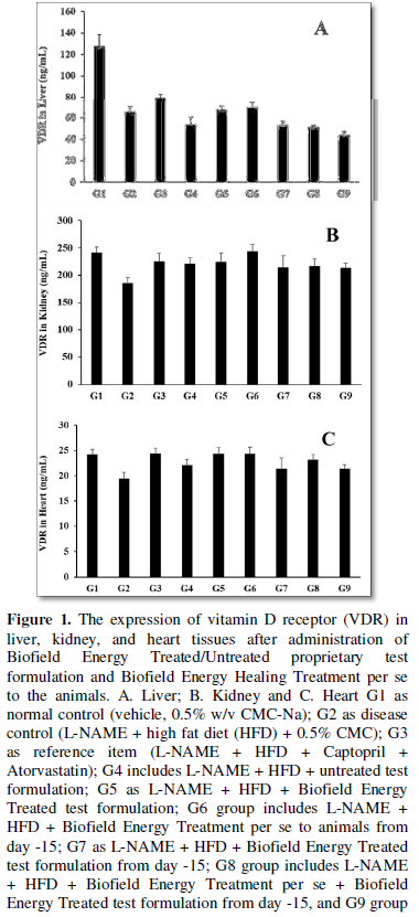
The expression of vitamin D receptor (VDR) in liver, kidney, and heart tissues after administration of Biofield Energy Treated/Untreated proprietary test formulation and Biofield Energy Healing Treatment per se to the animals are shown in Figure 1. In the liver tissue, the disease control (L-NAME + high fat diet (HFD) + 0.5% CMC) group (G2) showed the level of VDR as 65.09 ± 4.94 ng/mL, which was decreased by 48.93% as compared with the normal control (G1, 127.46 ± 11.02 ng/mL) group. However, the level of VDR expression was increased by 21.80% i.e., 79.28 ± 3.60 ng/mL in the positive control (captopril + atorvastatin) group (G3) in liver homogenate as compared to the G2 group. The level of VDR was increased by 2.45% and 6.48% in the G5 (L-NAME + HFD + the Biofield Energy Treated test formulation) and G6 (L-NAME + HFD + Biofield Energy Treatment per se to animals from day -15) groups, respectively with reference to G2 group. Further, the level of VDR expression was increased by 25.12% and 30.03% in the G5 and G6 groups, respectively with reference to untreated test formulation (G4) group (Figure 1A).
Moreover, in the kidney tissue, the disease control (L-NAME + high fat diet (HFD) + 0.5% CMC) group (G2) showed the level of VDR as 185.07 ± 10.31 ng/mL, which was decreased by 22.96% as compared with the normal control (G1, 240.23 ± 11.50 ng/mL) group. However, positive control (captopril + atorvastatin) treatment group (G3) the level of VDR in kidney homogenate was increased by 21.40% i.e. 224.67 ± 15.11 ng/mL as compared to the G2 group. The level of VDR was increased by 18.94%, 21.08%, 31.30%, 15.12%, 17.12%, and 15.06% in the G4 (L-NAME + HFD along with untreated test formulation), G5 (L-NAME + HFD + the Biofield Energy Treated test formulation) and G6 (L-NAME + HFD + Biofield Energy Treatment per se to animals from day -15), G7 (L-NAME + HFD + the Biofield Energy Treated test formulation from day -15), G8 (L-NAME + HFD + Biofield Energy Treatment per se + Biofield Energy Treated test formulation from day -15), and G9 (L-NAME + HFD + Biofield Energy Treatment per se animals + untreated test formulation) groups, respectively with reference to disease control (G2) group. Further, the level of VDR expression was increased by 1.80% and 10.390% in the G5 and G6 groups, respectively as compared to the untreated test formulation (G4) group (Figure 1B).
Additionally, in the heart tissue, the disease control (L-NAME + high fat diet (HFD) + 0.5% CMC) group (G2) showed the level of VDR as 19.37 ± 1.24 ng/mL, which was decreased by 19.56% as compared with the normal control (G1, 24.08 ± 1.12 ng/mL) group. However, positive control (captopril + atorvastatin) treatment group (G3) the level of VDR in kidney homogenate was increased by 25.59% i.e., 24.33 ± 1.14 ng/mL as compared to the G2 group. The level of VDR was increased by 13.64%, 25.23%, 25.45%, 9.99%, 19.42%, and 9.94% in the G4 (L-NAME + HFD + untreated test formulation), G5 (L-NAME + HFD + the Biofield Energy Treated test formulation) and G6 (L-NAME + HFD + Biofield Energy Treatment per se to animals from day -15), G7 (L-NAME + HFD + the Biofield Energy Treated test formulation from day -15), G8 (L-NAME + HFD + Biofield Energy Treatment per se + Biofield Energy Treated test formulation from day -15), and G9 (L-NAME + HFD + Biofield Energy Treatment per se animals + untreated test formulation) groups, respectively with reference to disease control (G2) group. Further, the level of VDR expression was increased by 10.21%, 10.40%, and 5.10% in the G5, G6, and G8 groups, respectively as compared to the untreated test formulation (G4) group (Figure 1C).
Numerous experimental studies indicate that VDR may play an important role in controlling blood pressure, cardiac hypertrophy, atherosclerosis, and heart failure [32]. It is also observed the usefulness in the prevention or treatment of cardiovascular disease [33]. In chronic kidney disease (CKD) deficiency of vitamin D is very common and cause various adverse effects [34]. All-inclusive, in this study the Biofield Energy Treated test formulation and Biofield Energy Treatment per se increased VDR expression in the vital organs like liver, kidney, and heart tissues that could be beneficial in the cardiovascular disorders.
Estimation of Telomerase Enzyme
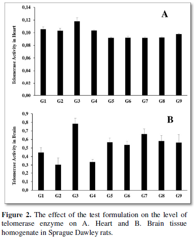
The levels of telomerase activity in heart and brain tissues were measured in all the experimental groups and the data are shown in Figure 2. The disease control (L-NAME + high fat diet, HFD + 0.5% CMC) group (G2) group showed the value of telomerase as 0.1 ± 0.00, which was decreased by 2.53% as compared with the normal control (G1, 0.11 ± 00) group in the heart tissue. While, the positive control (captopril + atorvastatin) treatment group (G3) increased the level of telomerase by 14.38% i.e., 0.12 ± 0.01 ng/mL as compared to the G2 group. The level of telomerase was altered in all the treatment groups as compared to both disease control (G2) and untreated test formulation group (G4) (Figure 2A).
Further, in the brain tissue, the level of telomerase in the disease control (L-NAME + high fat diet, HFD + 0.5% CMC) group (G2) was 0.3 ± 0.08, which was decreased by 31.93% as compared with the normal control (G1, 0.45 ± 0.06) group. While, the positive control (captopril + atorvastatin) treatment group (G3) increased the level of telomerase by 158.09% i.e., 0.78 ± 0.06 as compared to the G2 group. The level of telomerase in the brain tissue was significantly increased by 11.90%, 88.67%, 78.59%, 120.29%, 93.24%, and 87.63% in the G4 (L-NAME + HFD along with untreated test formulation), G5 (L-NAME + HFD + the Biofield Energy Treated test formulation) and G6 (L-NAME + HFD + Biofield Energy Treatment per se to animals from day -15), G7 (L-NAME + HFD + the Biofield Energy Treated test formulation from day -15), G8 (L-NAME + HFD + Biofield Energy Treatment per se plus the Biofield Energy Treated test formulation from day -15), and G9 (L-NAME + HFD + Biofield Energy Treatment per se animals plus the untreated test formulation) groups, respectively as compared to the disease control (G2) group. Further, the level of telomerase was increased by 66.47%, 57.58%, 94.38%, 70.50%, and 65.56% in the G5, G6, G7, G8, and G9 groups, respectively as compared to the untreated test formulation (G4) group (Figure 2B). Various clinical investigation reported that low telomerase activity and short leukocyte telomere length is responsible for the development of hypertension, atherosclerotic plaque, etc. [35-37]. However, in this study the Biofield Energy Treated test formulation and Biofield Energy Treatment per se has significantly increased the telomerase activity in brain tissue, which could be beneficial in the cardiac patients.
Estimation of citrate synthase enzyme in muscle
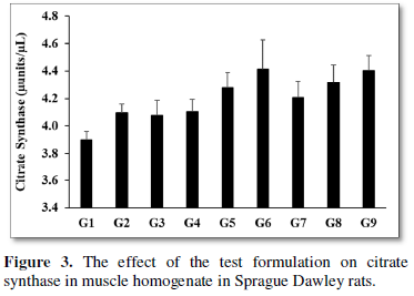
Citrate synthase enzyme was measured in the muscle tissue homogenate after treatment with the test formulation, and the data are shown in (Figure 3). The level of citrate synthase in the disease control (L-NAME + high fat diet (HFD) + 0.5% CMC) group (G2) was 4.10 ± 0.07 µunit/µL, and in the normal control group (G1, 3.90 ± 0.06 µunit/µL). Moreover, the positive control (captopril + atorvastatin) treatment (G3) showed the level of citrate synthase i.e., 4.08 ± 0.11 µunit/µL. The level of citrate synthase was increased by 4.31%, 7.65%, 2.62%, 5.14%, and 7.36% in the G5 (L-NAME + HFD + the Biofield Energy Treated test formulation), G6 (L-NAME + HFD + Biofield Energy Treatment per se to animals from day -15), G7 (L-NAME + HFD + the Biofield Energy Treated test formulation from day -15), G8 (L-NAME + HFD + Biofield Energy Treatment per se plus the Biofield Energy Treated test formulation from day -15), and G9 (L-NAME + HFD + Biofield Energy Treatment per se animals plus the untreated test formulation) groups, respectively, as compared to the both disease control group (G2) as well as the untreated test formulation group (G4) (Figure 3). Mitochondria play a vital role in skeletal muscles functions and metabolic health. Citrate synthase is a key mitochondrial enzyme used as a biomarker of mitochondrial content and function in mammals [38,39]. Overall, in this experiment the Biofield Energy Treated test formulation and Biofield Energy Treatment per se marginally increased the level of citrate synthase enzyme, which might be helpful for the management of cardiovascular disorders.
This experimental, four preventive maintenance groups were used. These groups were G6, G7, G8, and G9. Results showed the significant slowdown of cardiovascular-related symptoms/complications and reduced the chances of disease susceptibility. Based on the findings, it suggests that the Biofield Energy Healing Therapy/Blessing was found to be most effective and benefited to prevent and protect from the occurrence of any type of diseases and that will ultimately improve the overall health and quality of life in human.
CONCLUSION
The study findings showed that the expression of vitamin D receptor (VDR) in liver tissue was significantly increased by 25.12% and 30.03% in the G5 and G6 groups; respectively as compared to the (L-NAME + HFD + untreated test formulation) group (G4). Moreover, the expression of VDR in the kidney tissue was increased by 21.08%, 31.30%, 15.12%, 17.12%, and 15.06% in the G5, G6, G7, G8, and G9 groups, respectively as compared to the disease control (G2) group. Moreover, the level of VDR expression in the heart tissue was increased by 25.23%, 25.45%, and 19.42% in the G5, G6, and G8 groups, respectively as compared to the G2 group. Besides, the level of telomerase enzyme was significantly increased by 88.67%, 78.59%, 120.29%, 93.24%, and 87.63% in G5, G6, G7, G8, and G9 groups, respectively as compared to the G2 group. All-inclusive, the Biofield Energy Treated/Blessed test formulation and Biofield Energy Healing Treatment/Blessing (the Trivedi Effect®) per se showed fruitful results with respect to different tissue biomarkers in the preventive maintenance groups (G6, G7, G8, and G9) in L-NAME and High Fat Diet-Induced cardiovascular disorders rat model study. It also helped to slow down the cardiovascular disease progression and disease-related complications of the overall animal’s health. These data suggested that Biofield Energy Treatment per se and/or Biofield Energy Treated Test formulation in combination would be the best treatment strategies in order to prevent and protect from the occurrence of any type of diseases. Therefore, the Biofield Energy Treatment might act as a preventive maintenance therapy in order to maintain good health, or full restoration of health or improve the overall health and quality of life in human. This therapy might also reduce the severity of various type of acute/chronic diseases like auto-immune, inflammatory, and many thyroid disorders. Overall, the data suggested the Biofield Energy Treated/Blessed test formulation and Biofield Energy Treatment per se in showed significant action on thyroid gland with respect to biomarkers, as a CAM. This test formulation also can be used against fibromyalgia, Addison disease, multiple sclerosis, myasthenia gravis, rheumatoid arthritis, aplastic anemia, Crohn’s disease, psoriasis, chronic fatigue syndrome, vitiligo, and alopecia areata, dermatitis, ulcerative colitis, hepatitis, mental disorders, diverticulitis, Parkinson’s, and stroke in the improvement of overall health and quality of life.
ACKNOWLEDGEMENT
The authors are grateful to Dabur Research Foundation, Trivedi Science, Trivedi Global, Inc., and Trivedi Master Wellness for the assistance and support during the work.
- Szekely Y, Arbel Y (2018) A review of interleukin-1 in heart disease: Where do we stand today? Cardiol Ther 7(1): 25-44.
- WHO (2019) Cardiovascular diseases. Available online at: https://www.who.int/health-topics/cardiovascular-diseases/#tab=tab_1
- Raj K, Chanu SI, Sarkar S (2012) Decoding complexity of aging. Cell Dev Biol 1: e117.
- World Health Organization (2018) Interesting facts about ageing. Available online at: http://www.who.int/ageing/about/facts/en/
- Robinson WH, Lindstrom TM, Cheung RK, Sokolove J (2013) Mechanistic biomarkers for clinical decision making in rheumatic diseases. Nat Rev Rheumatol 9(5): 267-76.
- Chen S, Sun Y, Agrawal DK (2015) Vitamin D deficiency and essential hypertension. J Am Soc Hypertens 9(11): 885-901.
- Jih-Kai Yeh, Mei-Hsiu Lin, Chao-Yung Wang (2019) Telomeres as Therapeutic Targets in Heart Disease. J Am Coll Cardiol Basic Trans Sci 4(7): 855-865.
- Moslehi J, DePinho RA, Sahin E (2012) Telomeres and mitochondria in the aging heart. Circ Res 110(9): 1226‐1237.
- Mamdani F, Rollins B, Morgan L,Myers RM, Barchas JD, et al. (2015) Variable telomere length across post-mortem human brain regions and specific reduction in the hippocampus of major depressive disorder. Transl Psychiatry 5(9): e636.
- Larsen S, Nielsen J, Hansen CN, Nielsen LB,Wibrand F, et al. (2012) Biomarkers of mitochondrial content in skeletal muscle of healthy young human subjects. J Physiol 590(14): 3349-3360.
- Vigelsø A, Andersen NB, Dela F (2014) The relationship between skeletal muscle mitochondrial citrate synthase activity and whole-body oxygen uptake adaptations in response to exercise training. Int J Physiol Pathophysiol Pharmacol 6(2): 84-101.
- Byrne JH, Voogt M, Turner KM, Eyles DW, McGrath JJ, et al. (2013) The impact of adult vitamin D deficiency on behavior and brain function in male Sprague-Dawley rats. PLoS One 8(8): e71593.
- Rayman MP (2000) The importance of selenium to human health. Lancet 356: 233-241.
- Beard JL, Connor JR (2003) Iron status and neural functioning. Ann Rev Nutr 23: 41-58.
- Peres FF, Lima AC, Hallak JEC, Crippa JA, Silva RH, et al. (2018) Cannabidiol as a promising strategy to treat and prevent movement disorders? Front Pharmacol 9: 482.
- Nagarkatti P, Pandey R, Rieder SA, Hegde VL, Nagarkatti M (2009) Cannabinoids as novel anti-inflammatory drugs. Future Med Chem 1(7): 1333-1349.
- Kang S, Min H (2012) Ginseng, the 'Immunity Boost': The effects of Panax ginseng on immune system. J Ginseng Res 36(4): 354-368.
- Maizes V, Rakel D, Niemiec C (2009) Integrative medicine and patient-centered care. Explore (NY) 5(5): 277-289.
- Bischof M, Del Giudice E (2013) Communication and the emergence of collective behavior in living organisms: A quantum approach. Mol Biol Int 2013: 987549.
- Cassidy CM (2004) What does it mean to practice an energy medicine? J Altern Complement Med 10(1): 79-81.
- Barnes PM, Bloom B, Nahin RL (2008) Complementary and alternative medicine use among adults and children: United States, 2007. Natl Health Stat Report 12: 1-23.
- Fan K wai (2005) National Center for Complementary and Alternative Medicine Website. J Med Libr Assoc 93: 410-412.
- Wisneski L, Anderson L (2009) The Scientific Basis of Integrative Medicine. Boca Raton, FL: CRC Press pp: 205.
- Trivedi MK, Branton A, Trivedi D, Jana S (2021) Isotopic abundance ratio analysis of consciousness energy healing treated folic acid. Food Nutr Current Res 4(2): 290-295.
- Trivedi MK, Branton A, Trivedi D, Nayak G, Mondal SC, et al. (2015) Morphological characterization, quality, yield and DNA fingerprinting of biofield energy treated alphonso mango (Mangifera indica). J Food Nutri Sci 3: 245-250.
- Trivedi MK, Jana S (2019) In vitro assessment of the biofield treated test item on cardiac function using rat cardiomyocytes cell line (H9c2) via multiparametric analysis. J Hypertension Cardiol 2(4): 1-12.
- Trivedi MK, Branton A, Trivedi D, Jana S (2021) Effect of consciousness energy healing treatment on the metal profile and properties of tellurium. Eng Technol Open Acc 3(5): 555623.
- Mahendra KT, Alice B, Dahryn T, Snehasis J (2021) Consciousness energy healing treatment impacted the isotopic abundance ratio of 6-Mercaptopurine (6-MP). Nov Appro Drug Des Dev 5(5): 555673.
- Trivedi MK, Jana S (2021) Anti-aging activity of biofield energy treated novel proprietary test formulation by assessment of vital biomarkers in cerebrospinal fluid (CSF) in Sprague Dawley rats. On J Neur Br Disord 5(2): 2021.
- Trivedi MK, Jana S (2021) Evaluation of biofield energy healing treatment based proprietary test formulation on gut health potential in colon cancer cell line (HT-29). J Pharmacol Clin Res 8(4): 555743.
- Trivedi MK, Branton A, Trivedi D, Jana S (2020) The consciousness energy healing treatment and its impact on the isotopic abundance ratio analysis of flutamide. Drug Des Int Prop Int J 3(5): 2020.
- Judd SE, Tangpricha V (2009) Vitamin D deficiency and risk for cardiovascular disease. Am J Med Sci 338(1): 40‐44.
- Gardner DG, Chen S, Glenn DJ (2013) Vitamin D and the heart. Am J Physiol Regul Integr Comp Physiol 305(9): R969‐R 977.
- Gonzalez-Parra E, Rojas-Rivera J, Tuñón J, Praga M, Ortiz A, et al. (2012) Vitamin D receptor activation and cardiovascular disease. Nephrol Dial Transplant 27 (4): 17-21.
- Yeh JK, Wang CY (2016) Telomeres and telomerase in cardiovascular diseases. Genes (Basel) 7(9): 58.
- Zurek M, Altschmied J, Kohlgrüber S, Ale-Agha N, Haendeler J (2016) Role of telomerase in the cardiovascular system. Genes (Basel) 7(6): 29.
- Zhan Y, Hägg S (2019) Telomere length and cardiovascular disease risk. Curr Opin Cardiol 34(3): 270-274.
- Vaanholt LM, Al-Tarrah M, Gray SR, Speakman JR, Hambly C, et al. (2019) Low citrate synthase activity is associated with glucose intolerance and lipotoxicity. J Nutr Metab 2019: 8594825.
- Mukherjee A, Smitherman TC, Robinson JB Jr, Butsch RW, Richards EG, et al. (1980) Studies on human heart citrate synthase. Adv Myocardiol 1: 329-337.


QUICK LINKS
- SUBMIT MANUSCRIPT
- RECOMMEND THE JOURNAL
-
SUBSCRIBE FOR ALERTS
RELATED JOURNALS
- Journal of Psychiatry and Psychology Research (ISSN:2640-6136)
- International Journal of Radiography Imaging & Radiation Therapy (ISSN:2642-0392)
- Journal of Allergy Research (ISSN:2642-326X)
- Journal of Neurosurgery Imaging and Techniques (ISSN:2473-1943)
- Journal of Oral Health and Dentistry (ISSN: 2638-499X)
- Journal of Ageing and Restorative Medicine (ISSN:2637-7403)
- Journal of Otolaryngology and Neurotology Research(ISSN:2641-6956)


